Retrophotos. Physics.
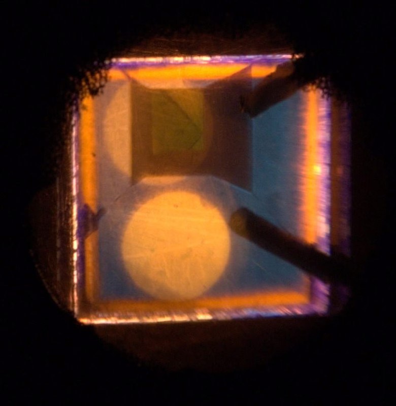
It’s a photo from my previous life as a physicist. To be honest, it’s one of the greatest surprises of my life. You take a glass-clear piece of diamond—perfectly transparent and homogeneous. You put it in the electron microscope, close the lid, pour liquid nitrogen into the vacuum pumps, and wait four hours. Then you start and tune the electron-beam system, cool the sample holder with liquid nitrogen, adjust the optical system—and then… you see this picture. It’s a natural diamond, and the growth sectors are clearly visible. You can see blue, orange, and green lines of luminescence.
Blue region — N3 center (λ ≈ 415 nm), an aggregated-nitrogen defect.
Green — H3 center, formed by irradiation + annealing (often enhanced by plastic deformation).
Yellow — NV⁰ center at 575 nm (nitrogen + vacancy).
The electron microscope was half of the setup. The other half was a fairly large spectrometer. We recorded spectra in different areas of the samples and tried to capture the diffusion of vacancies.
Those days gave me the habit of writing down everything you do in your experiments, very carefully. When you're writing, everything feels obvious. A month later, it's anything but obvious—and you curse that guy who didn’t put in enough effort to write down the crucial details you now crave while trying to write an article.